Hadrontherapy is a very promising treatment technique that proposes to replace the traditional photon or electron beams with proton beams, more specifically called proton therapy, or ions ranging from helium to oxygen
The Normandy region has decided to create the first hadrontherapy research center in France. This is the ARCHADE project, which should deliver its first beams around 2025.
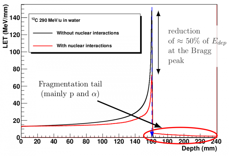
The undeniable advantage of ion beams, compared to photon or electron beams, is that each incident particle is relatively undeflected by the constant Coulombic interactions that it undergoes during its slowing down. In other words, an ion goes (almost) straight ahead and stops precisely where you want it to (by adjusting its initial energy). This is a ballistic advantage that all ions share. This ballistic advantage is coupled with a dosimetric advantage linked to the nature of the coulombic interaction. The slower the ion, the more it is slowed down, and the more efficiently its energy is transferred to the surrounding medium. The dosimetric advantage is materialized by the appearance of a dose peak at the end of the ion’s path. This is the Bragg peak. The Bragg peak is the manifestation of the Linear Energy Transfer, or LET, of the ions on the medium. We will see later that dosimetry and ballistics are coupled to a biological benefit, this time specific to the incident ion.
Transfert d'énergie linéique en fonction du parcours pour une ion 12C dans l'eau (Simulation GEANT4). La courbe noire correspond à une simulation dans laquelle le processus de collisions entre noyaux a été désactivé.
However, the incident ions do not only undergo coulombic interactions. Indeed, they may collide with an atom of the medium through which they pass (the patient in this case). This type of event is all the more likely when the incident ion or the target have large apparent surfaces. This probability is measured by a quantity called the effective collision cross section which depends on the incident ion and the target. To give an idea, approximately 50% of the ions in a carbon beam undergo this kind of collision over a 16cm path in the patient.
This nuclear collision is violent and leads to the dislocation of the incident ion and the atom in fragments of random nature. This is the fragmentation phenomenon. The fragments of the beam (lighter) go globally at the speed of the incident ion. As they are lighter, they are slowed down less efficiently and go beyond the expected arrival point of the incident ion, thus delocalizing the dose beyond the tumor. These fragments go further the smaller their size. This is the fragmentation tail. In summary, for a target located at 15 cm depth, approximately 50% of the ions sent will fragment the fragments and contribute to the fragmentation tail. This represents approximately 10 to 20% of the delivered dose, but this dose is delocalized downstream of the tumor. It is therefore important to know the nature and physical parameters of these fragments (nature, charge, angle, energy). This knowledge is summarized in the differential effective fragmentation cross section. The effective fragmentation cross sections are directly used in treatment planning software to simulate the dose deposition in the patient.
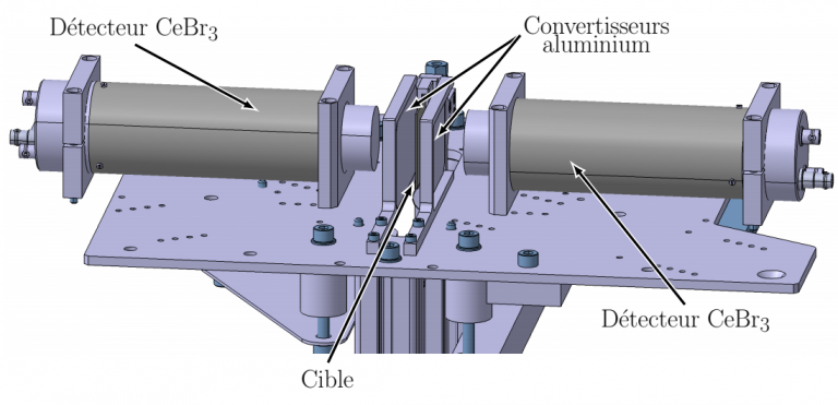
In addition to the basic phenomenon (how the dose is deposited in the patient), these effective sections also have another interest. Indeed, among the fragments produced (particularly on the target), there are β+ emitting radioisotopes that are exploitable in positron emission tomography (PET). The tomography of these emitters then allows the qualitative verification of the dose deposition in the patient. This is referred to as on-line PET imaging. Experimental campaigns using the PEPIT measurement device have taken place in order to measure the production cross sections of these β+ emitters, the most recent of which dates from spring 2019.
Accomplishment:
GrAMI has a significant activity in measuring these differential fragmentation cross sections. Four low-energy measurement campaigns have been performed at GANIL with carbon ions since 2011. The measured cross sections are available on the WEB site http://hadrontherapy-data.in2p3.fr.
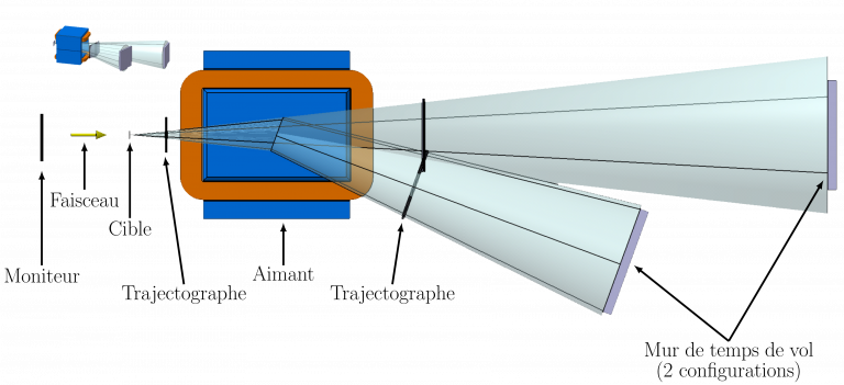
Schéma du spectromètre de masse FRACAS permettant la mesure des sections efficaces de fragmentation des particules du faisceau.
A dedicated measurement device named FRACAS (FRAgmentation du CArbone et Sections efficaces) is under construction to perform these measurements of effective sections of fragmentation of the beam at high energy at the ARCHADE center.
The PEPIT device is dedicated to the measurement of β+ emitters in the target during irradiation by ion beams.
Dessin du dispositif PEPIT permettant la mesure des sections efficaces de production des émetteurs β+ après l’irradiation d’une cible